As the site is updated, each listing includes the shipping cost. Some listings which I have not updated still give you calculated shipping costs based on weight and size of package. (In the sections I have updated) If you select several different listings, we will consolidate your order and charge you the actual cost of the entire package. The shipping over charge will be refunded to you, when your order is shipped.
Mother of Pearl
Nacre also known as mother of pearl, is an organic–inorganic composite material produced by some molluscs as an inner shell layer. It is also the material of which pearls are composed. It is strong, resilient, and iridescent.
Nacre is found in some of the most ancient lineages of bivalves, gastropods, and cephalopods. The inner layer in the great majority of mollusk shells are porcellaneous, not nacreous, and this usually results in a non-iridescent shine, or more rarely in non-nacreous iridescence such as flame structure as is found in conch pearls.
The outer layer of cultured pearls and the inside layer of pearl oyster and freshwater pearl mussel shells are made of nacre. Other mollusk families that have a nacreous inner shell layer include marine gastropods such as the Haliotidae, the Trochidae and the Turbinidae.
Nacre is composed of hexagonal platelets of aragonite (a form of calcium carbonate) 10–20 µm (micrometers) wide and 0.5 µm (micrometers) thick arranged in a continuous parallel lamina. Depending on the species, the shape of the tablets differs; in Pinna, the tablets are rectangular, with symmetric sectors more or less soluble(REF:Nudelman, Fabio; Gotliv, Bat Ami; Addadi, Lia; Weiner, Steve (2006). "Mollusk shell formation: Mapping the distribution of organic matrix components underlying a single aragonitic tablet in nacre". Journal of Structural Biology). Whatever the shape of the tablets, the smallest units they contain are irregular rounded granules. These layers are separated by sheets of organic matrix (interfaces) composed of elastic biopolymers (such as chitin, lustrin and silk-like proteins).
(REF: Cuif J.P. Dauphin Y., Sorauf J.E. (2011). Biominerals and fossils through time. Cambridge: Cambridge University Press)
Nacre appears iridescent because the thickness of the aragonite platelets is close to the wavelength of visible light. These structures interfere constructively and destructively with different wavelengths of light at different viewing angles, creating structural colors.
The crystallographic c-axis points approximately perpendicular to the shell wall, but the direction of the other axes varies between groups. Adjacent tablets have been shown to have dramatically different c-axis orientation, generally randomly oriented within ~20° of vertical. In bivalves and cephalopods, the b-axis points in the direction of shell growth, whereas in the monoplacophora it is the a-axis that is this way inclined.
(REF: Metzler, Rebecca; Abrecht, Mike; Olabisi, Ronke; Ariosa, Daniel; Johnson, Christopher; Frazer, Bradley; Coppersmith, Susan; Gilbert, PUPA (2007). "Architecture of columnar nacre, and implications for its formation mechanism". Physical Review Letters) (REF:Olson, Ian; Kozdon, Reinhard; Valley, John; Gilbert, PUPA (2012). "Mollusk shell nacre ultrastructure correlates with environmental temperature and pressure". Journal of the American Chemical Society. ) (REF: Checa, Antonio G.; Ramírez-Rico, Joaquín; González-Segura, Alicia; Sánchez-Navas, Antonio (2008). "Nacre and false nacre (foliated aragonite) in extant monoplacophorans (=Tryblidiida: Mollusca)". Naturwissenschaften)
The process of how nacre is formed is not completely clear. It's been observed in a species called Pinna nobilis (the Pin Shell), where it starts as tiny particles (~50–80 nm) grouping together inside a natural material. These particles line up in a way that resembles fibers, and they continue to multiply. When there are enough particles, they come together to form early stages of nacre. The growth of nacre is regulated by organic substances that determine how and when the nacre crystals start and develop.
(REF: Jackson, D. J.; McDougall, C.; Woodcroft, B.; Moase, P.; Rose, R. A.; Kube, M.; Reinhardt, R.; Rokhsar, D. S.; et al. (2009). "Parallel Evolution of Nacre Building Gene Sets in Molluscs". Molecular Biology and Evolution)Each crystal, which can be thought of as a "brick," is thought to rapidly grow to match the full height of the layer of nacre. They continue to grow until they meet the surrounding bricks. This produces the hexagonal close-packing characteristic of nacre. The growth of these bricks can be initiated in various ways such as from randomly scattered elements within the organic layer, well-defined arrangements of proteins, or they may expand from mineral bridges coming from the layer underneath.
(REF: Checa, Antonio G.; Ramírez-Rico, Joaquín; González-Segura, Alicia; Sánchez-Navas, Antonio (2008). "Nacre and false nacre (foliated aragonite) in extant monoplacophorans (=Tryblidiida: Mollusca)". Naturwissenschaften. 96) (REF: Addadi, Lia; Joester, Derk; Nudelman, Fabio; Weiner, Steve (2006). "Mollusk Shell Formation: A Source of New Concepts for Understanding Biomineralization Processes". ChemInform. 37) (REF: Nudelman, Fabio; Gotliv, Bat Ami; Addadi, Lia; Weiner, Steve (2006). "Mollusk shell formation: Mapping the distribution of organic matrix components underlying a single aragonitic tablet in nacre". Journal of Structural Biology. 153 )(REF:Schäffer, Tilman; Ionescu-Zanetti, Cristian; Proksch, Roger; Fritz, Monika; Walters, Deron; Almquist, Nils; Zaremba, Charlotte; Belcher, Angela; Smith, Bettye; Stucky, Galen (1997). "Does abalone nacre form by heteroepitaxial nucleation or by growth through mineral bridges?". Chemistry of Materials. 9)(REF: Checa, Antonio; Cartwright, Julyan; Willinger, Marc-Georg (2011). "Mineral bridges in nacre". Journal of Structural Biology. 176)
What sets nacre apart from fibrous aragonite, a similarly formed but brittle mineral, is the speed at which it grows in a certain direction (roughly perpendicular to the shell). This growth is slow in nacre, but fast in fibrous aragonite. (REF Bruce Runnegar & S Bengtson. Origin of Hard Parts — Early Skeletal Fossils )
A 2021 paper in Nature Physics examined nacre from Unio pictorum, noting that in each case the initial layers of nacre laid down by the organism contained spiral defects. Defects that spiraled in opposite directions creating distortions in the material that drew them towards each other as the layers built up until they merged and cancelled each other out. Later layers of nacre were found to be uniform and ordered in structure.[21][28]
(REF: Beliaev, N.; Zöllner, D.; Pacureanu, A.; Zaslansky, P.; Zlotnikov, I. (2021). "Dynamics of topological defects and structural synchronization in a forming periodic tissue". Nature Physics. 124 )(REF: Meyers, Catherine (January 11, 2021). "How Mollusks Make Tough, Shimmering Shells". Inside Science)
Nacre is secreted by the epithelial cells of the mantle tissue of various mollusks. The nacre is continuously deposited onto the inner surface of the shell, the iridescent nacreous layer, commonly known as mother of pearl. The layers of nacre smooth the shell surface and help defend the soft tissues against parasites and damaging debris by entombing them in successive layers of nacre, forming either a blister pearl attached to the interior of the shell, or a free pearl within the mantle tissues. The process is called encystation and it continues as long as the mollusk lives.

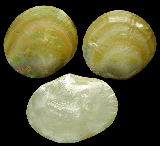
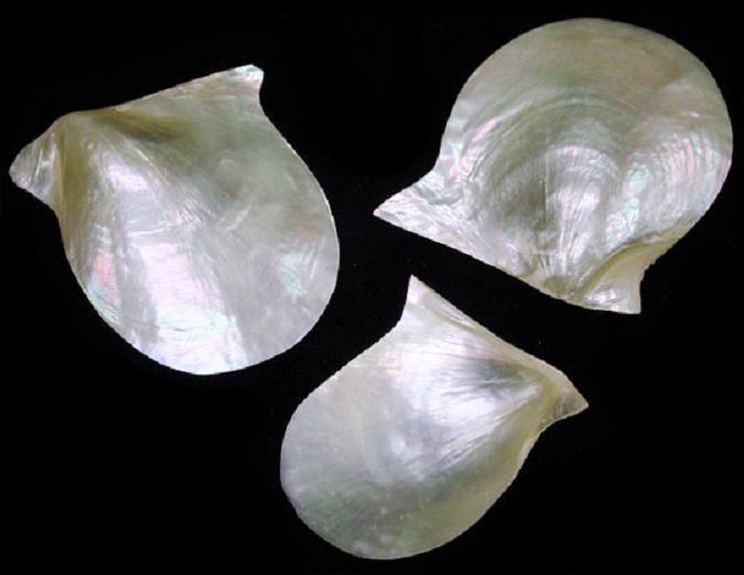
Gold Mother of Pearl Plate
B1-15
One Polished Gold Mother of Pearl Plate, measuring 5 to 5 1/4 inches. ..... $18.25
B2-15
One polished gold Mother of Pearl plate, measuring approximately 5.5 inches.......$22.45
B3-15
One polished gold Mother of Pearl plate, measuring approximately 6 inches....... $23.95

Mother of Pearl Natural
X1-15
One natural Mother of Pearl, measuring 2 to 3 inches...... .30